- Home
- Ankle and Foot
- Overpronation
- Navicular Drop Test
Navicular Drop Test
The height of the navicular bone sitting versus standing. It is the simplest measurement of how much the arch gives way under the body's own weight, and it takes four minutes.
- Gear
- A bare foot, a pen, a ruler, and a chair to sit on.
- Stance
- Sit with the foot on the floor, knee bent to 90 degrees, and mark the height of the navicular tubercle on the floor with a pen line.
- Duration
- 180 s
- Camera angle
- frontal, lens level with the landmark
- Position
- Measure the height of the navicular from the floor while sitting, then stand up and measure it again on the same spot. The difference between the two is the drop.
Bone landmarks
-
Navicular tuberosity — coordinate id
navicular -
Lateral malleolus — coordinate id
lateral-malleolus -
Calcaneus — coordinate id
calcaneus
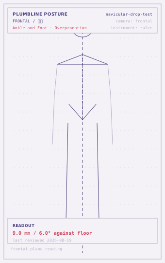
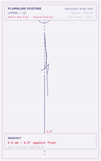
Within range
0-5 mmCriterion: The navicular height drops 5 mm or less between sitting and standing, and the medial arch is still visible from the front when standing.
The arch is bearing the body's weight without collapsing. Re-check when footwear or training volume changes.
Borderline
6-10 mmCriterion: A drop of 6 to 10 mm, or a drop that is more than 3 mm larger on one side than the other measured the same way.
The arch has control in the sitting position and loses part of it under load. This is the band where a change of shoe or insole is worth measuring rather than guessing at.
Deviated
over 10 mmCriterion: More than 10 mm of drop, or the navicular reaching the floor, or the drop coming with pain along the arch or the plantar surface of the heel.
A large drop with pain is not a posture finding. Have it examined, and do not start arch exercises until it has been.
| Tier | What to do |
|---|---|
| Low | Reading sits in the within-range band and is symmetric side to side: re-measure every 8–12 weeks and record which way the number is moving. |
| Moderate | Reading sits in the borderline band, or the two sides differ clearly: re-measure every 4–6 weeks and log the daily trigger (hours seated, one-sided load). Change the situation before adding anything to the routine. |
| High | Reading sits in the deviated band, or several screens at the same station all read deviated: get a clinician to rule out a structural or neural cause before continuing to self-screen. |
| Referral threshold | Arch pain that persists at rest, or that wakes the person at night, or that comes with numbness in the foot, needs assessment. |
Commonly co-occurring
How to read this
One bone, two positions
The arch is a structure that holds a shape, and the only interesting question about it is how much of that shape survives the person’s own body weight. Measuring the arch’s height while sitting answers nothing; measuring it again while standing answers everything.
The navicular is the landmark because it is the highest point of the arch’s inner border and it is easy to find by touch. Its height above the floor sitting and standing is the standard way of putting a number on a flexible flat foot.
Procedure
Sit on a chair with the foot flat on the floor and the knee bent to ninety degrees. Find the navicular and measure its height above the floor, or mark the height on a wall with a pencil and measure the mark.
Then stand up on the same spot, in the same foot position, and measure again. Subtract.
Do both feet, and write down both numbers plus the difference.
Reading it
Five millimetres or less of drop, with the arch still visible from the front when standing, is the reference. Six to ten millimetres means the arch has control sitting and loses part of it loaded.
More than ten millimetres, or a navicular that reaches the floor, or a drop that comes with pain under the arch or the heel, is a clinical picture rather than a postural one. Have it looked at before beginning any arch work.
What the number is for
The drop is most useful as a comparison. Between sides, it identifies a foot that has changed relative to the other one. Over time, it shows whether a change of footwear, or a change in how much time is spent standing, has actually done anything — which is the question people answer by feel, usually wrongly.
Questions
Where exactly is the navicular?
The bony bump on the inside of the foot, roughly a thumb's width forward of and below the inner ankle bone. Press along the inner border until you find the most prominent point; it is usually easy to identify once the foot is relaxed.
Why measure sitting first?
Because sitting is the position where the arch is not being asked to do anything. The difference between the two positions is the part of the arch's height that depends on load, which is the part that changes with shoes and training.
Does a big drop always mean a problem?
No. A mobile, flexible foot that drops a long way is common and frequently painless. The drop becomes interesting when it comes with symptoms, or when it is clearly larger on one side than the other.
Disclaimer
This page is informational only. It is not a medical diagnosis and it is not treatment or rehabilitation advice. Run every self-screen inside a pain-free range; stop and see a clinician for pain, numbness, weakness, night pain, or anything that started after an injury. The readout criteria are drawn from healthy-adult norms — prior injury, surgery and individual variation all shift the numbers.
lastReviewed 2026-08-19 · record navicular-drop-test · published 2026-08-27