- Home
- Head and Neck
- Forward Head
- Craniovertebral Angle from a Phone Photo
Craniovertebral Angle from a Phone Photo
The angle between the tragus-to-C7 line and the horizontal, read from a single lateral photograph. One number, one frame, one camera distance held constant.
- Gear
- The same lateral photograph used by the tragus angle screen, plus a protractor or angle app.
- Stance
- Stand relaxed, side-on, gaze level. No correction and no instruction — the frame has to capture the position you are not thinking about.
- Duration
- 90 s
- Camera angle
- frontal, lens level with the landmark
- Position
- Mark the tragus and C7 on the image and measure the angle of that line above the horizontal. The craniovertebral angle is the complement of the angle the same line makes with vertical.
Bone landmarks
-
Tragus — coordinate id
tragus -
C7 spinous process — coordinate id
c7 -
Vertex — coordinate id
vertex
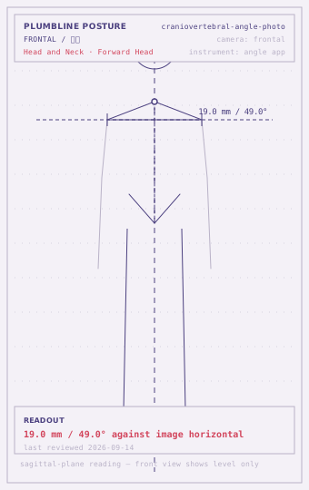
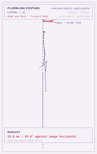
Within range
55-70 °Criterion: The tragus-to-C7 line sits between 55 and 70 degrees above the horizontal in the resting frame.
This is the range reported for asymptomatic adults in the measurement literature. Take the single number as a baseline and re-photograph in three months.
Borderline
45-54 °Criterion: The angle sits between 45 and 54 degrees, or the resting frame and the retracted frame differ by less than 6 degrees.
A smaller craniovertebral angle on its own is not a finding; a small angle combined with a small cued change is the pattern worth logging and re-checking in four weeks.
Deviated
under 45 °Criterion: Under 45 degrees, or the cued frame shows no change at all, or the shoulder sits forward of the hip line in the same frame.
Below this value the measurement is describing the whole upper quadrant rather than the neck alone. Read the thoracic and shoulder position from the same frame before drawing a conclusion.
| Tier | What to do |
|---|---|
| Low | Reading sits in the within-range band and is symmetric side to side: re-measure every 8–12 weeks and record which way the number is moving. |
| Moderate | Reading sits in the borderline band, or the two sides differ clearly: re-measure every 4–6 weeks and log the daily trigger (hours seated, one-sided load). Change the situation before adding anything to the routine. |
| High | Reading sits in the deviated band, or several screens at the same station all read deviated: get a clinician to rule out a structural or neural cause before continuing to self-screen. |
| Referral threshold | A clinician should see any neck symptom that comes with arm pain, hand numbness, unsteadiness on the feet, or that began after a collision. |
Commonly co-occurring
How to read this
One angle, held constant
The craniovertebral angle is the most commonly reported single number in forward-head measurement, and it earns that position by being simple: mark the tragus, mark C7, measure the angle of the line above the horizontal. Larger means the head is more nearly stacked; smaller means it is further forward.
What the number cannot do is stand alone. The published ranges move with age, with sex, and with the population sampled, and the difference between a “normal” and a “deviated” value can be smaller than the error introduced by moving the camera half a metre. This screen is therefore written to be used as a series, not as a verdict. One photograph tells you nothing. Five photographs across a year, all taken the same way, tell you whether anything is moving.
Keeping the series comparable
Three things must stay fixed or the series is worthless:
- Camera distance and height. Mark the floor where the tripod stands, and note the height. Perspective distortion changes the measured angle, and it does so differently at different distances.
- The frame you take it in. Half-body lateral is the practical choice: it includes C7 and the tragus with room to spare. A full-body frame shrinks both landmarks to a few pixels.
- The time of day. Write it in the record. A resting position at 08:00 and one at 19:00 are different measurements of different states, not two samples of the same thing.
Reading it against the same photograph’s other angles
The single most useful thing about a lateral photograph is that it contains more than one measurement. In the same frame you can mark the acromion and the greater trochanter and read whether the shoulder is forward of the hip. You can mark the ASIS and PSIS and read the pelvic tilt. When the craniovertebral angle is low, the question that decides what to do next is whether the frame shows a head problem or a whole-spine problem, and that question is answerable from the same image without taking another one.
Where the number stops
A low craniovertebral angle is a geometric statement. It is not a pain diagnosis, it does not predict who will develop symptoms, and the studies that report the ranges do not support using one value as a threshold for treatment. Record it, track it, and treat a change of less than about five degrees as indistinguishable from measurement noise unless the series is long enough to show a trend.
Questions
Why is a larger angle better here?
A larger craniovertebral angle means the head is closer to being stacked over C7. As the head moves forward, the line from C7 up to the tragus flattens toward the horizontal and the angle shrinks. So a change downward means the head has moved further forward, not that the neck has improved.
My phone app measures from vertical, not horizontal. Does that break the test?
No. The two are complements — a 50-degree craniovertebral angle above horizontal is a 40-degree deviation from vertical. What matters is that you use the same one every time; mixing conventions between sessions turns a real change into an apparent one.
Should I take the photo in the morning or evening?
Pick one and stay with it. The resting head position after eight hours of desk work is measurably different from the position on waking, so a morning photo compared against an evening one will show a change that is about the time of day. State the time in your record.
Disclaimer
This page is informational only. It is not a medical diagnosis and it is not treatment or rehabilitation advice. Run every self-screen inside a pain-free range; stop and see a clinician for pain, numbness, weakness, night pain, or anything that started after an injury. The readout criteria are drawn from healthy-adult norms — prior injury, surgery and individual variation all shift the numbers.
lastReviewed 2026-09-14 · record craniovertebral-angle-photo · published 2026-09-20